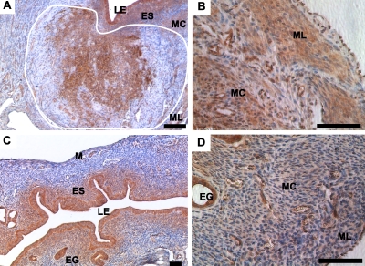
FIG. 3.
FRAP1 expression in Amhr2tm3(cre)Bhr;Ctnnb1tm1Mmt uteri. A) FRAP1 expression was detected by immunohistochemistry at high levels in the central areas of a leiomyoma (outlined in white) in a mutant uterus, the epithelial cells, the ES as well as in the circular myometrial layer (MC) and longitudinal myometrial layer (ML). B) A higher magnification view of FRAP1 expression in the MC and ML of a mutant mouse. C) In control uteri, strong FRAP1 expression is found in the EG, LE, and ES. D) Expression in the control myometrium appears limited to the endothelial cells. LE, luminal epithelium; ES, endometrial stroma; M, myometrium; EG, endometrial glands. Bars = 50 μm (A and C), 100 μm (B and D).

