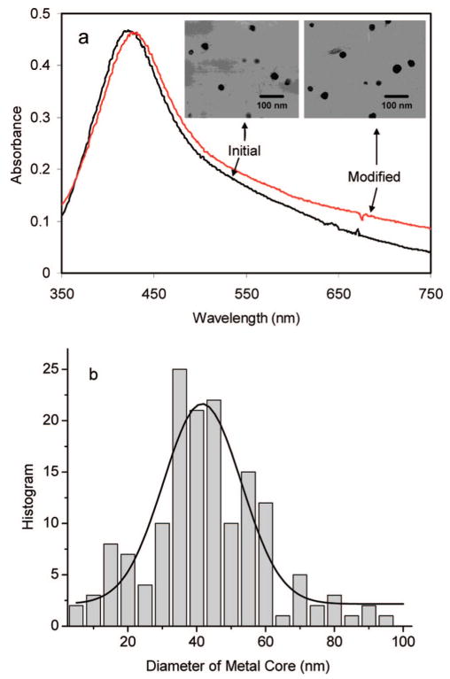
Figure 1.
(a) Absorbance spectra of initial tiopronin-coated silver particles and aminated metal particles with 40 nm metal cores. Their transmission electron micrographs (TEM) images are given in the insets a and b, respectively. (b) Histogram of size distribution for the tiopronin-coated silver particles achieved from TEM images. At least 200 metal particles were counted from the TEM images.

