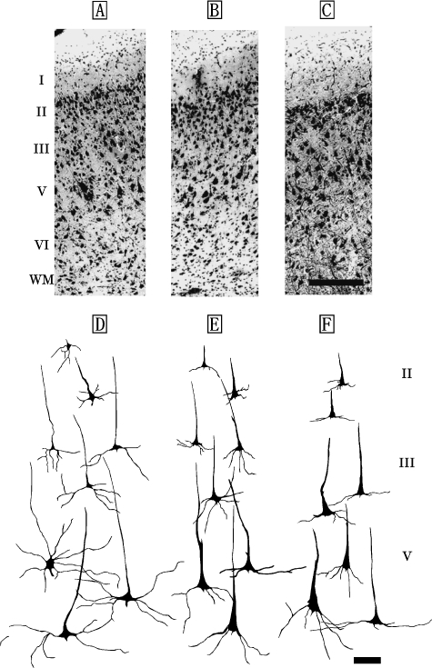
Fig. 1.
Photomicrographs of Nissl-stained, coronal sections of primary motor area (M1) in the Risso's dolphin (A), striped dolphin (B), and bottlenose dolphin (C). Scale bar indicates 300µm (A–C). Schematic drawings represent Golgi-impregnated neurons of layers II, III, and V in M1 of the Risso's dolphin (D), striped dolphin (E) and bottlenose dolphin (F). Scale bar indicates 100µm (D–F). I: layer I (molecular layer); II: layer II (external granular layer); III: layer III (external pyramidal layer); V: layer V (internal pyramidal layer); VI: layer VI (fusiform layer); WM: white matter.

