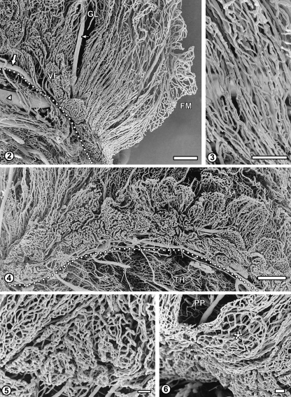
Fig. 2.
The vascular pattern of the posterior part of the left ventricle plexus. This area is characterized by a dense array of straight, parallel blood vessels: arterioles, venules and capillaries which form tubular plexuses around bigger vessels. These vessels blend with irregular vascular network of the glomus (GL). In the narrow zone of villous fringe (VF) along the attached margin (dotted line), capillaries form relatively small, rounded or elongated baskets, which become bigger and more leaf-like as the fringe continues anteriorly. Flat multiple capillary arcades of the free margin (FM) are also seen. Large arrow: medial branch of anterior choroidal artery; small arrow: its branch; arrowhead: choroido-ventricular vein. Bar=1000µm.
Fig. 3 Sheath-like capillary networks surrounding parallel arterioles and venules in the posterior part of the choroid plexus. Bar=500µm.
Fig. 4 The C-shaped villous fringe of the right ventricle plexus composed of basket-like or leaf-like capillary plexuses of the choroid villi. The elongated villi emerge from behind and are extending dorsally over the basket-like structures. Dotted line indicates attached margin of the plexus. Anterior-posterior axis: right-to-left. TH, thalamus. Bar=1000µm.
Fig. 5 Higher magnification of the fringe posterior–anterior transition area (left-to-right). Small, nearly flat and very dense basket-like capillary networks located under the posterior part of the plexus become bigger, looser and more folded, being separated by narrow sulci. Bar=100µm.
Fig. 6 Fragment of the fringe under the posterior part of the plexus (PP): a flat, discoid capillary plexus (asterisk). Bar=100µm.

