Fig. 1.
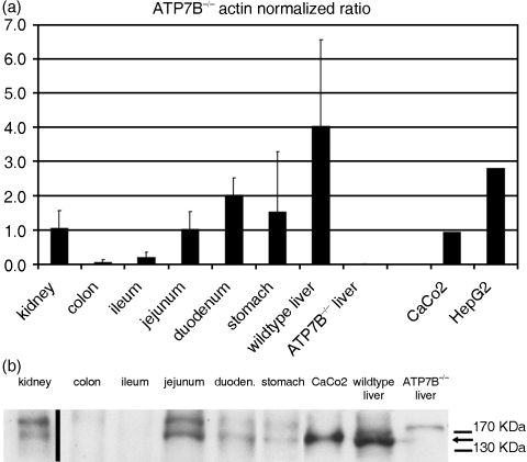
a) Real-time PCR quantification of ATP7B in murine tissues and human CaCo-2 cells. Mouse tissues were evaluated for their mRNA content of mATP7B. The results confirmed a strong expression of mATP7B in liver and moderate expression in kidney, stomach, duodenum and jejunum. Tissue samples derived from wild-type colon, wild-type ileum or liver of ATP7B−/– mice did not show a significant signal above background (*). Error bars indicate the standard deviation of three independent experiments (different mice). Caco-2 and HepG2 cells were evaluated for their mRNA content of hATP7B/ WDP and showed a significant expression (right columns). Arbitrary units were obtained by normalizing the signal to the internal control (values shown are 10−3 * signal ATP7B/signal actin). b) Immunoblot analysis of ATP7B in murine tissues and CaCo-2 cells. Tissues or cells were lysed, and 25µg of protein was separated by SDS-PAGE, transferred to nitrocellulose, incubated with the affinity-purified ATP7B antibody, and analysed by chemiluminescence. The lane with mouse kidney lysate is derived from a different blot.

