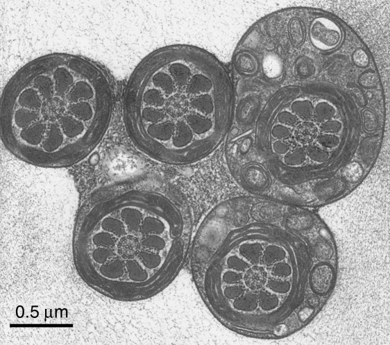
Fig. 3.
A group of sperm flagella sectioned at the level of the midpiece, from a thin section cut perpendicularly to the freezing plane. At this level the flagella are stuck together by the granular material, which has been shown to lie also between the sperm heads (Fig. 1). This mechanical coupling must have a role in the flagellar synchronization – which is generally explained in the literature on cilia and flagella as being due to viscous coupling. Again, the structure of the midpiece and of the cytoplasmic droplet (top right) is well preserved and conventional. Scale bar=0.5µm.

