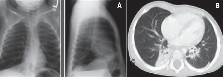
Figure 2.

A patient with Spastic diplegia, brain atrophy, respiratory syncytial virus infection, gastroesophageal reflux and bilateral bronchiectasis. A- Chest X-ray: Increase density in lower lobes bilaterally. B. CT chest: Dilatation of bronchi of both lower lobes with collapse/ consolidation of affected lobes.
