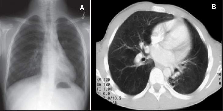
Figure 5.

A patient with TB infection proved by lung biopsy and sputum culture and LLL bronchiectasis. A. Chest C-ray: Left lower lobe showing evidence of airway disease with bronchiectasis, bronchial wall thickening and minor peribronchial infiltrate. B. CT chest: Bronchiectatic changes in the left lower lobe, superior and anterior segments. Some areas of fibrosis with contraction of the left lung.
