
Figure 1. SDS-polyacrylamide gel electrophoresis.
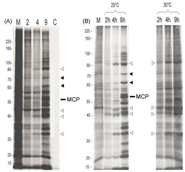
Panel A: As indicated in Methods and Materials, confluent monolayers of FHM cells were either mock-infected (M, lane 1) or infected with FV3 and radiolabeled with [35S]methionine from 1-2, 2-4, or 7-9 hr post infection (lanes 2 – 4). At the indicated times, replicate cultures were lysed and viral proteins were separated on 10% SDS polyacrylamide gels. One set of FV3-infected cultures was incubated in the presence of 100 μg/ml cycloheximide (C, lane 5) and radiolabeled as described above from 7 – 9 hr p.i. Panel B: A FV3 temperature-sensitive mutant defective in viral DNA synthesis (ts5) was used to infect FHM cells at permissive (25°C) and non-permissive (30°C) tempera tures. Replicate cultures were radiolabeled from 1 – 2, 2 – 4, and 7 – 9 hr p.i., and visualized by autoradiography after gel electrophoresis. Mol wt markers are shown to the left of panels A and B. The position of the MCP is marked with an arrow, and representative early and late viral proteins are indicated by open (early) and filled (late) triangles, respectively.