
Figure 1.
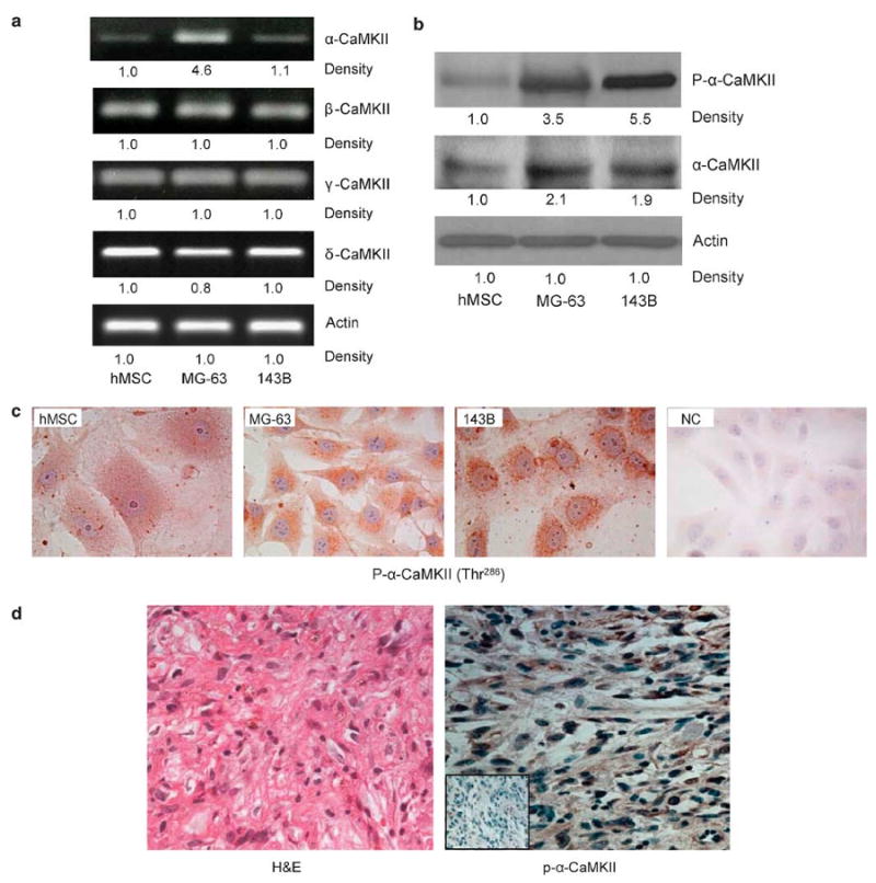
Human osteosarcoma expresses high levels of α-CaMKII. MG-63,143B and hMSC cells were used. Representative images from three experiments of (a) RT-PCR using CaMKII isoforms or actin-specific primers. (b) Western blots using antibodies directed against phosphorylated and total α-CaMKII and actin (c) Immunohistochemistry staining using a specific antibody directed against p-α-CaMKII. (d) Primary human osteosarcoma tissue was surgically removed, formalin-fixed and paraffin-embedded. H&E staining (left) shows osteosarcoma. Immunohistochemical staining (right) was performed using an antibody against p-α-CaMKII (brown), counterstained with hematoxylin (blue). NC, without immunoreactivity, is shown in the low left inset. Photomicrographs were obtained at × 400 magnification, and are representative of the four different patients.