
Figure 5.
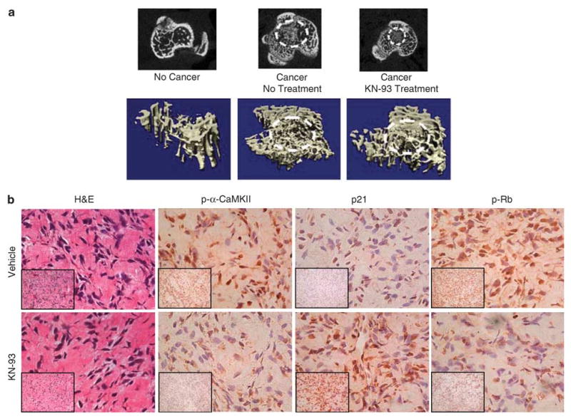
The CaMKII antagonist, KN-93, decreases the in vivo growth of human osteosarcoma. MG-63 cells were subcutaneously or intratibially injected into 5-week-old male athymic nu/nu mice that were divided into untreated and KN-93-treated groups. Mice were treated every other day for 6 weeks. (a) After mice were killed, tibiae were removed, formalin fixed and MicoCT scanning was performed. The upper panel shows a two-dimensional section in the proximal tibia, where osteosarcoma has grown (indicated by broken white circle), whereas the lower panel shows a 3D image of the cross-sectional proximal tibia also showing the tumor. (b) Subcutaneous tumors were removed, formalin fixed and paraffin embedded. H&E and IHC staining were performed for p21, p-Rb and p-α-CaMKII. All images were obtained at × 200 (lower left insets) and × 400. The results showed no change in the cytomorphology comparing the control to the KN-93-treated tumors, but upregulation of p21 and downregulation of p-α-CaMKII and p-pRb, as assessed by IHC.