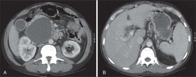
Figure 1.
A Contrast-enhanced computed tomography scan of the upper abdomen. A relatively thin-walled cystic mass is identified in the region of the pancreatic head measuring 9.6 cm × 8.5 cm × 8.4 cm in size. Multiple punctate pancreatic calcifications are seen, consistent with a history of pancreatitis. B The cyst indents the common bile duct anteriorly, with intrahepatic biliary dilation

