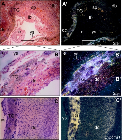
Figure 1.
Localization of Star and Cyp11a1 mRNAs in E9.5 mouse placenta. By d 9.5 mouse pregnancy, implantation sites were prepared for in situ hybridization as described in Materials and Methods. Shown are low and high-power bright-field micrographs depicting a sagittal-to-embryo/transverse-to-uterus section stained with hematoxylin and eosin (A and C) and dark-field micrographs of the same microscope field (A′ and C′). A and A′ note a strong hybridization signal of Star transcript in the TG cells, but no labeling signals in the decidua basalis (db), the placental layers of spongiotrophoblasts (sp) and labyrinth (lb), the embryo (e) and the yolk sac (ys). B and B′, Bright- and dark-field micrographs, respectively, of the boxed area in A and A′ (magnification, ×40). Note a strong labeling of the TG cells, in contrast to lack of signal in the embryo and the yolk sac cells. A weak signal is observed in the decidua capsularis (dc). C and C′, Bright- and dark-field micrographs, respectively, of a decidua capsularis (dc) section processed for Cyp11a1 in situ hybridization, similar to that shown in B/B′ (magnification, ×33). Note a strong labeling of the TG cells, in contrast to the lack of signal in the yolk sac cells (ys). A weak signal is observed in the decidua capsularis (dc), as described before (24). n, Nucleus.

