
Figure 2. Morphological analysis of differentiated hES cells.
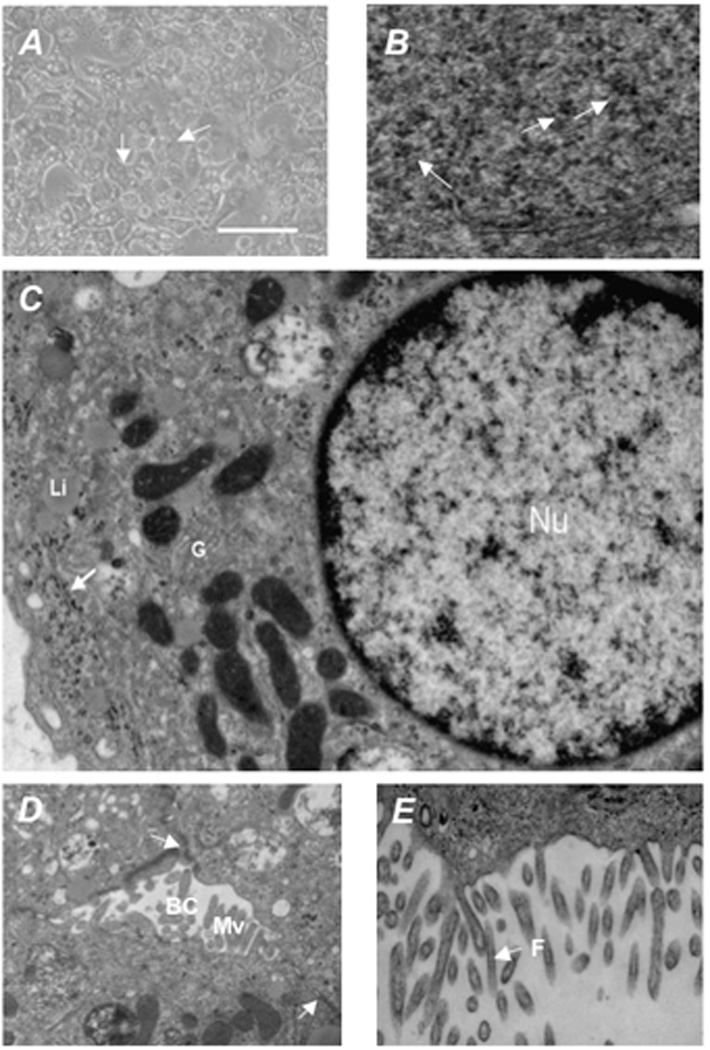
Morphological analysis of differentiated hES cells in culture indicate that the differentiation program generated cells morphologically similar to hepatocytes, being polygonal in shape with multiple nuclei. (A) Transmission electron microscopy of differentiated hES showed (B) accumulation of glycogen rosettes (arrows), (C) round nuclei (Nu) with evenly distributed chromatin, Golgi complexes (G), and liposomes (Li), with glycogen rosettes identified again by an arrow, and (D, E) well developed bile canaliculi (BC) with apical microvilli (Mv) containing filaments (F) , tight junctions (arrows). Scale bar = 50 µm. Original magnification × 30,000.