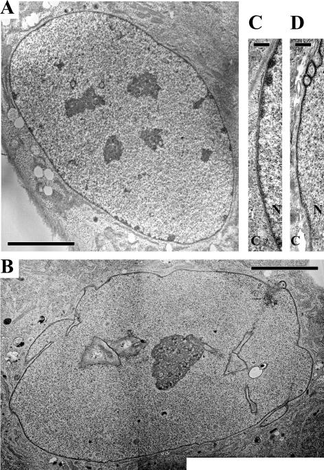
Figure 3.
Electron microscopic observation of normal and HGPS fibroblasts. Low-magnification views show peripheral heterochromatin and nucleoplasmic heterochromatic foci in the normal nucleus (A), which are both absent in the highly lobulated HGPS nucleus (B). A high-magnification view of the nuclear envelope of a HGPS nucleus (D) shows a loss of peripheral heterochromatin and a thickening of the nuclear lamina compared with a normal nucleus (C). (C) Cytoplasm; (N) nucleus. Bars: A,B, 5 μM; C,D, 300 nm.

