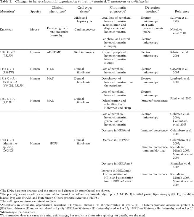
Table 1.
Changes in heterochromatin organization caused by lamin A/C mutations or deficiencies
aThe DNA base pair changes and the amino acid changes (in parentheses) are shown.
bThe phenotypes are as follows: autosomal-dominant Emery-Dreifuss muscular dystrophy (AD-EDMD), familial partial lipodystrophy (FPLD), mandibuloacral dysplasia (MAD), and Hutchinson-Gilford progeria syndrome (HGPS).
cThe cell types or tissue examined are listed.
dAlterations in chromatin organization described. (H3K9me3) Histone H3 thrimethylated at Lys 9; (HP1) heterochromatin-associated protein 1; (H3K9me1) histone H3 monomethylated at Lys 9; (H3K27me3) histone H3 thrimethylated at Lys 27; (H4K20me3) histone H4 thrimethylated at Lys 20.
eMicroscopic methods used.
fThis mutation does not cause an amino acid change, but results in alternative splicing (for details, see the text).

