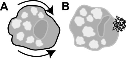
Figure 1.
Surface receptor capping in Entamoeba histolytica. Cartoons show amoeba cells. (A) Once antigens on the amoeba surface are recognized by host immune components (indicated by black line), they are rapidly translocated toward the posterior pole (arrows). (B) At the posterior end, membranes harboring surface receptors form into a complex array of vesicles termed the uroid prior to being shed (Silva et al. 1975).

