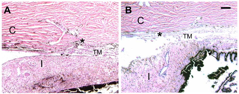
Figure 1.
Hematoxlyn-eosin stained anterior chamber section of cat eyes. A. Control (non-laser treated) eyes displaying histological structure of the anterior segment. B. A representative anterior eye section that underwent selective laser trabeculoplasty (SLT). No significant structural changes were found between control and treated trabecular meshwork. The location of the Cornea (C), Iris (I) and Trabecular meshwork (TM) are as indicated. (*) identifies Schlemm’s canal (SC), Bar = 100 μm.

