
Figure 1.
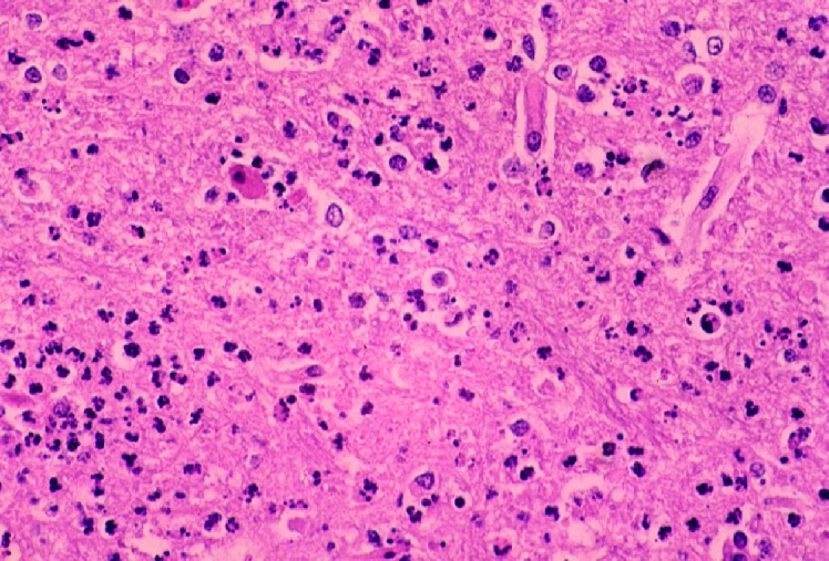
Photomicrograph of a section of the cerebral cortex from horse with Eastern equine encephalomyelitis virus infection. Note the dense neutrophilic response, vascular damage, and fibrin thrombi. Hematoxylin and eosine stain.
Official websites use .gov
A
.gov website belongs to an official
government organization in the United States.
Secure .gov websites use HTTPS
A lock (
) or https:// means you've safely
connected to the .gov website. Share sensitive
information only on official, secure websites.
Photomicrograph of a section of the cerebral cortex from horse with Eastern equine encephalomyelitis virus infection. Note the dense neutrophilic response, vascular damage, and fibrin thrombi. Hematoxylin and eosine stain.