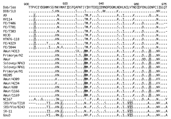
Figure 4.

Multiple alignment of partial deduced amino acid sequences of G2 region of hantaviruses. Amino acid sequences analyzed by using ClustalX (ver. 1.8) program. Amino acid positions indicated above sequences based on Haantan 76–118. First line shows the deduced amino acid of Dobrova/Saarema. Dots represent amino acids that are identical to those at corresponding positions in Dobrova/Saarema sequence. Amino acids that differ from those in the sequence are indicated at relevant positions. Hyphens are used in areas where amino acid sequence is not available. Signature amino acids are shaded.
