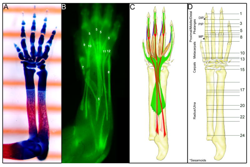
Fig. 1.

The tendons of the forelimb at E18.5.
(A) A ventral view of a skeletal prep of a forelimb from an embryo at E18.5 captured over a ruler showing 1mm gradation marks.
(B) A dorsal view of a skinned forelimb of an E18.5 ScxGFP embryo. The extensor tendons are identified with a number that identifies them in the tendon table (Table 1).
(C) Schematic drawing of the major flexor tendons in the forelimb at E18.5. Green – Flexor Digitorium Profundus tendon; Red – Flexor digitorium Sublimis tendon; Blue – Lumbrical muscles and tendons.
(D) A schematic drawing of the ventral side of the forelimb that serves to illustrate the position of sections in Figs. 2&3. DIP – Distal interphalangeal joint; PIP – proximal interphalangeal joint; MP – Metacarpophalangeal joint.
