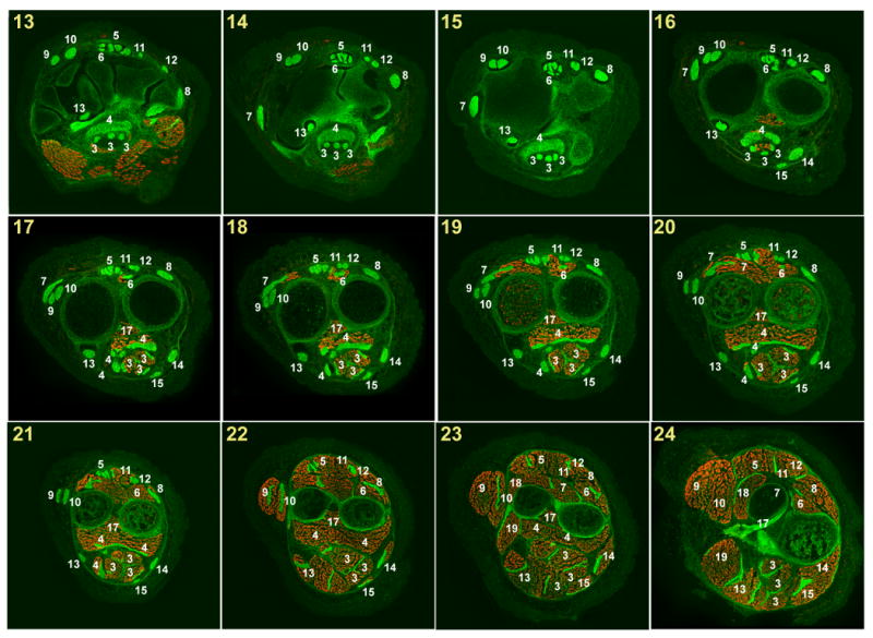
Fig. 3.

Muscles and tendons of the forelimb at E18.5 (2).
Successive cross sections of a forelimb from an E18.5 ScxGFP embryo stained for MHC. In all panels dorsal is up and anterior is to the left.
Panel numbers indicate the position of each section in the illustration in Fig. 1D.
White numerals – Tendon or muscle number in Table 1.
Red numerals – Ligament number in Table 2.
Red S – sesamoid bone.
