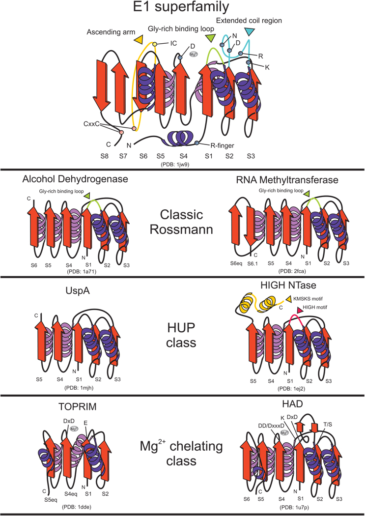
Figure 2. Topology diagrams and comparison with other Rossmann-like proteins.
Representative cores of Rossmann-like domains belonging to different classes of the fold are depicted as cartoons. Inserts and other lineage-specific features are depicted and labeled with various other colors. Gray spheres represent the magnesium ions in various active sites. Residues experimentally shown to contribute to catalytic activity in the given representatives are labeled. Strand numbers are given at the bottom of each strand; “eq” refers to strands that are spatially equivalent to strands in the E1 superfamily.

