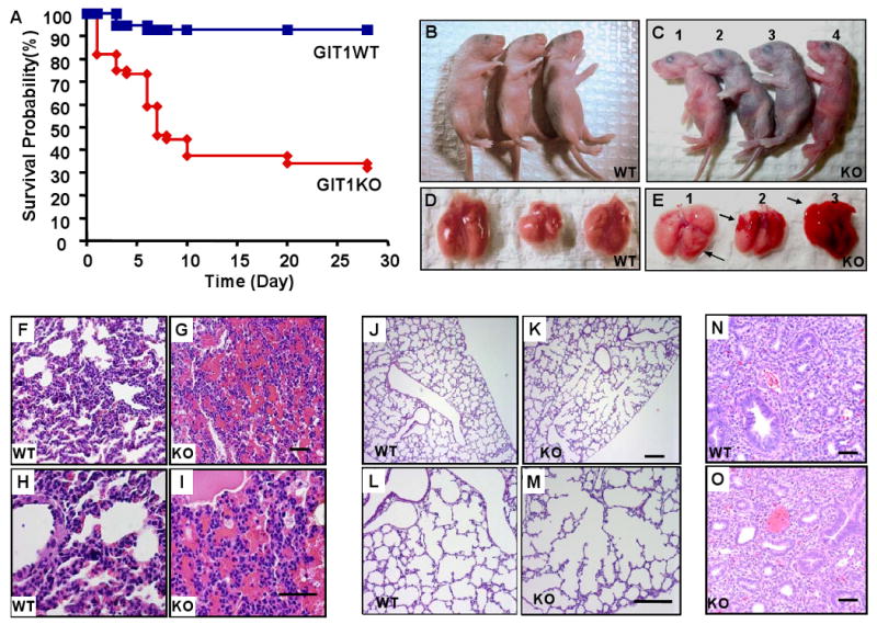
Figure 2. Survival, gross anatomy and histopathology in GIT1 WT and KO mice.

A. Kaplan-Meier survival analysis. B-C. GIT1 WT and KO neonates (P4): WT neonates (B) and two KO neonates appeared normal (C, #1 and 4) while others (C, #2 and 3) showed varying degrees of cyanosis and respiratory distress. D-E. Fetal lungs (P4) from WT mice (D) had a normal gross appearance, whereas lungs from KO mice (E) showed scattered hemorrhages. F-I. Hematoxylin and eosin (H&E) staining of lung sections from P5 GIT1 WT and KO mice. There was obvious hemorrhage in parenchyma of GIT1 KO mice (G, I) compared to WT mice (F, H). J-M. GIT1 WT and KO mice lungs were perfused as described in methods, then the tissues were embedded and stained with H&E. GIT1 WT mice show normal, well-developed saccular and alveolar airway structures (J, L), whereas GIT1 KO showed abnormally large airspaces (K; M). N-O H&E staining of lung sections from embryos (E14.5) of GIT1 WT and KO mice. There were no obvious differences in histological appearance between WT and KO embryonic lungs.
