
Figure 5. GIT1 is required for activation of MEK1, ERK1/2 and PLCγ signaling pathways in EC and lung.
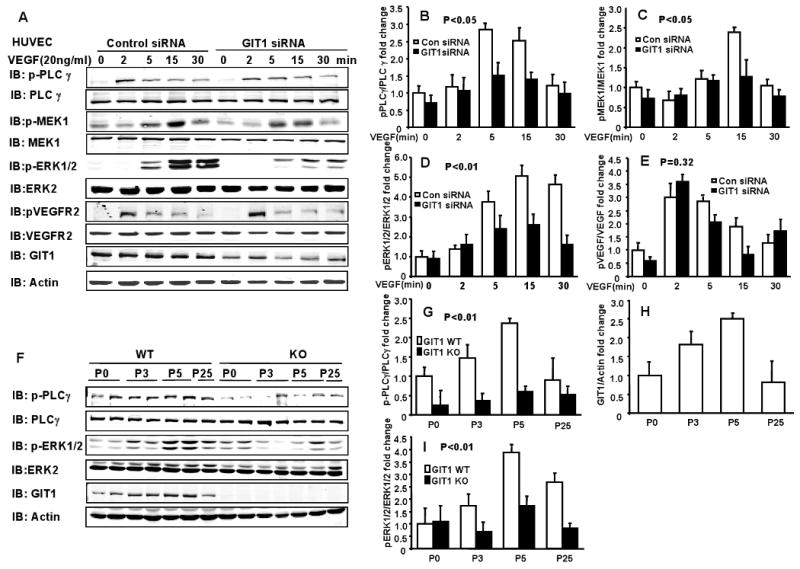
A. GIT1 and control siRNA were transfected for 48 h into HUVECs. After serum starvation for 4 h, the cells were stimulated with 20 ng/ml VEGF for the indicated times and phosphorylation of the indicated proteins was determined. B-E. Right panels are quantitation of relative increase of PLCγ, MEK1, ERK1/2 and VEGFR2 phosphorylation compared to control siRNA group without VEGF stimulation. P< 0.05 compared with control siRNA groups (mean ±SE; n =3). F. Phosphorylation of PLCγ and ERK1/2 as well as expression of GIT1 in lungs of GIT1 WT and KO mice were detected by immunoblot. Total PLCγ, ERK1/2 and actin were assayed as loading controls. G-I. Quantitation of relative changes of PLCγ and ERK1/2 phosphorylation, as well as GIT1 expression (normalized to actin in WT P0 group). P< 0.01 compared with WT (mean ±SE; n =4).