Fig. 2.
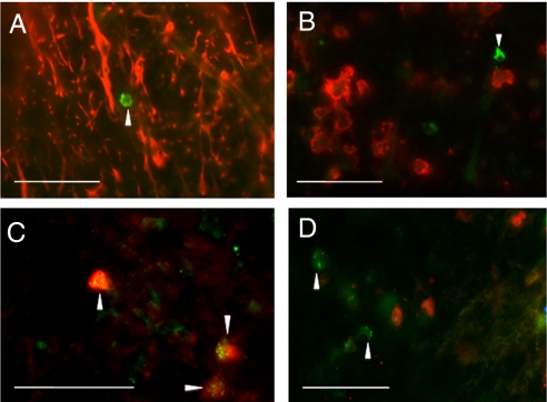
Galanin expression in the mouse spinal cord after EAE. Galanin-expressing cells (green) are indicated by arrowheads in all images. Galanin staining is not observed in (A) GFAP-positive astrocytes (red) or (B) CD11b-positive microglia (red). (C) Many PLPdsRED-positive oligodendrocytes express galanin, but (D) not in naive healthy (no EAE induction) spinal cord from PLPdsRED transgenic mice. (Scale bars, 50 μm.)

