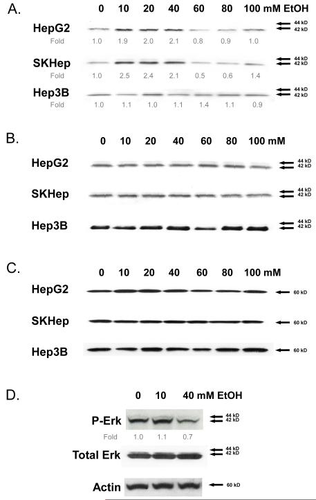
Fig. 1.
ERK activation in human hepatocellular carcinoma (HCC) cell lines (Hep3B, SKhep, and HepG2) following treatment with ethanol (0-100 mM) for 24 hours. Cell lysates were analyzed by Western immunoblotting to detect (A) phosphorylated ERK1,2 - fold expression relative to β-actin averaged from three independent experiments is shown; (B) total ERK1,2; and (C) β-actin. (D) Human hepatocytes treated with ethanol (0-40 mM) for 24 hours. Phosphorylated ERK1,2 with fold expression relative to β-actin, total ERK, and β-actin are shown. Representative Western immunoblots are presented; similar results were obtained in at least two independent experiments.

