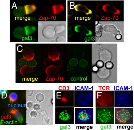
Fig. 2.
Galectin-3 is recruited to the IS. (A) Galectin-3-transfected (Gal3+) Jurkat T cells were stimulated for 5 min with SEE-pulsed RPMI 8866 B cells. The cells were processed for immunofluorescence staining of galectin-3. (B and C) Gal3+ Jurkat cells were stimulated with anti-CD3-coated latex beads and stained with anti-galectin-3 (B) or control antibody (C). (D) Activated mouse CD4+ T cells from wild-type mice were stimulated with anti-CD3-coated beads and the cells were stained for galectin-3. (E) Gal3+ Jurkat cells were stimulated to form the IS on supported lipid bilayers preloaded with fluorescence-labeled anti-CD3 and ICAM-1. The cells were stained for galectin-3. (F) Gal3+/+OTII CD4+ T cells were mixed with fluorescence-labeled anti-TCRβ and injected into flow cells coated with lipid bilayers preloaded with pMHC and fluorescence-labeled ICAM-1 to form the IS. The cells were stained for galectin-3.

