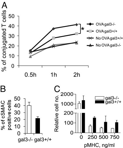
Fig. 3.
Galectin-3 inhibits cSMAC formation and destabilizes T-cell binding to APCs. (A) DiO-labeled gal3−/− and gal3+/+ OTII CD4+ T cells were mixed with DiI-labeled BMDC in the presence of 1 μg/mL OVA 323–339 peptide. The double fluorescence-labeled cell conjugates were detected by flow cytometry at the indicated time points. The percentages of cell conjugates were calculated by dividing the double-fluorescence labeled cell counts over the total stained T-cell counts. ANOVA, P < 0.05. (B) Gal3−/− and gal3+/+ CD4+ T cells were stimulated as mentioned in Fig. 2F and the ratios of the cSMAC-positive cell numbers over the total cell numbers were calculated. P < 0.01. (C) Gal3−/− and gal3+/+OTII CD4+ T cells were placed on the upper chamber of a transwell device on top of a membrane coated with ICAM-1 and pMHC. Cells migrated to the lower chambers were enumerated by flow cytometry. ANOVA, P < 0.0001.

