Fig. 6.
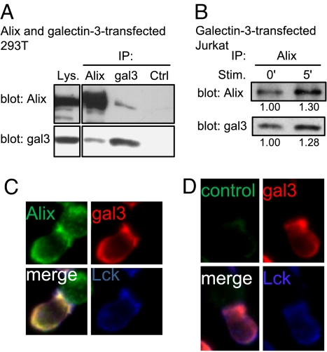
Galectin-3 is associated with Alix at the IS. (A) HEK-293T cells were co-transfected with plasmid DNAs containing Alix and galectin-3. The cells were treated with DSP and lysed, and the lysates were subjected to immunoprecipitation using anti-Alix, anti-galectin-3 (gal3) or control sera (Ctrl). The lysates (Lys.) and precipitates were immunoblotted with anti-Alix and anti-galectin-3 antibodies. (B) Gal3+ Jurkat cells were stimulated with anti-CD3-coated beads for 0 and 5 min and subjected to DSP cross-linkage. The lysates were immunoprecipitated with anti-Alix and immunoblotted with anti-Alix and anti-galectin-3 antibodies. The amounts of Alix and galectin-3 induced by the stimulus, relative to their baseline levels, are shown under each blot. (C and D) Galectin-3-transfected Jurkat T cells were stimulated for 5 min with SEE-pulsed RPMI 8866 B cells. The cells were fixed and stained with antibodies for galectin-3 (red) and Lck (blue), together with either anti-Alix antibody (green) (C) or a control antibody (D).

