Fig. 5.
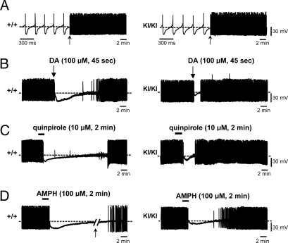
Decreased sensitivity to inhibition of firing induced by DA, quinpirole, and AMPH in R1441C KI nigral neurons. (A) Sample traces showing the comparable spontaneous, rhythmic action potentials recorded in DA neurons from +/+ (Left) and KI/KI (Right) mice. Firing activity is shown at different time scales marked by an arrow. (B) In midbrain slices from +/+ mice (left trace), bath-application of DA (100 μM, 45 s) hyperpolarizes the cell membrane and blocks the firing activity. On DA washout, the membrane potential slowly recovers, and the action potential discharge returns to control levels. DA-induced firing cessation is significantly shorter in nigral neurons of KI/KI mice (right trace). (C and D) Sample traces showing the reduced responses in firing of nigral neurons in KI/KI mice to bath-application of quinpirole (10 μM, 2 min) (C) or AMPH (100 μM, 2 min) (D). The arrow in D indicates a 10-min interruption in the trace of +/+ nigral neurons.

