Fig. 2.
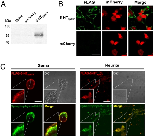
Subcellular localization of 5-HTapAC1. (A and B) Expression of FLAG-5-HTapAC1 in HEK293T cell. Expression was confirmed by western blot (A) and immunocytochemistry (B). In immunocytochemistry, FLAG-5-HTapAC1 (green) localized in the cytoplasmic membrane. mCherry-N1 (red) was co-transfected as a expression marker and diffusely distributed in the cytosol. (C) Co-localization of overexpressed FLAG-5-HTapAC1 (red) and synaptophysin-EGFP (green) in Aplysia sensory cells co-cultured with LFS motor neurons. Insets show three fold magnification images. FLAG-5-HTapAC1 and synaptophysin-EGFP are highly co-localized at synaptic varicosities, and partially co-localized at neurites and the plasma membrane, but not co-localized at the cytosol. (Scale bars, 30 μm.)

