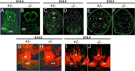
Fig. 5.
Staining of embryonic heads for phosphohistone H3 and β-gal. (A–F) Coronal sections stained for phosphohistone H3 (green), which is specific for mitotic cells. At E14.5 (A and B), E16.5 (C and D), and E18.5 (E and F), the number of cells containing the phosphorylated histone is decreased by 30–50% in bnc2−/− embryos. This diminution is particularly evident in the palatal processes at E14.5 (arrowheads). Notice in F the diminished size of the head of the homozygous mutant at E18.5. (G–J) Double-staining of palatal region for the phosphohistone and the β-gal (red). At E14.5 in the heterozygote, (G) the frequency of cells containing the phosphohistone is similar in areas expressing and not expressing bnc2lacZ. In the bnc2−/− embryo (H), the frequency of phosphohistone-containing cells is predominantly reduced in areas with high expression of the fusion gene. At E16.5 (I and J), the scarcity of mitoses in regions expressing the fusion gene is also observed, particularly in the nasal septum. m: mouth, md: mandible, n: nasal region, nc: nasal cavity, ns: nasal septum, pp: palatal processes, t: tongue.

