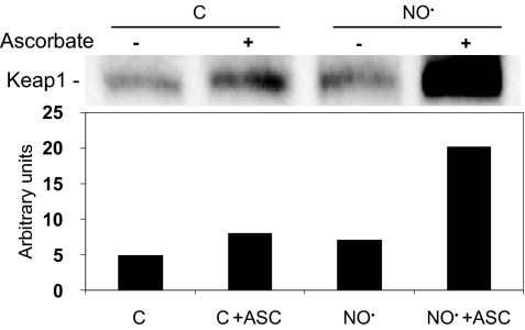
Fig. 7.
(S)-nitrosation of Keap1. HCT116 cells were exposed to 1,600 μM·min •NO and submitted to biotin switch assay of protein (S)-nitrosation as described in Materials and Methods. Biotinylated proteins were isolated and analyzed by Western blot with anti-Keap1 antibody. An increased signal in the presence of ascorbate (ASC), which displaces •NO from nitrosothiols, enhancing biotin labeling, verifies specificity of (S)-nitrosation. Cells exposed to argon were used as controls. The bar graph represents the densitometric analysis of Keap1 bands in the blot.

