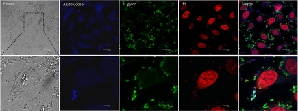
Fig. 1.
H. pylori obtains l-fucose from host cell membranes. Capan 1 cells were incubated with 200 μM tetra-O-acetyl-6-azido-l-fucose for 72 h, intensively washed 5 times with PBS, and then infected with H. pylori for 4 h. Cells were fixed, labeled with 1,8-naphthalimide alkyne (blue), stained with H. pylori-specific antibody (Alexa Fluor 488, green) and the nuclei-specific dye propidium iodide (PI, red), and examined by confocal fluorescence microscopy. Overlay appears as light green. (Upper scale bar, 20 μm. Lower scale bar, 5 μm.)

