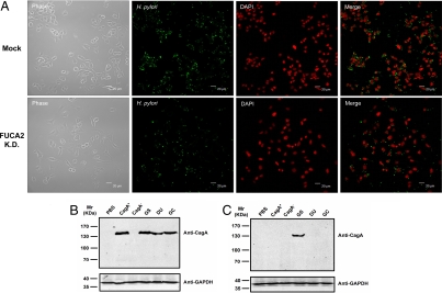
Fig. 3.
FUCA2 is essential for H. pylori adhesion to host cells. (A) Both mock-transfected Capan 1 cells and Capan 1-FUCA2 K.D. cells were infected with a comparable number of H. pylori, and doubly stained with anti-H. pylori (Alexa Fluor 488, green) and a nuclei-specific dye (DAPI, red). After 8 h of co-culturing, the number of adherent H. pylori was reduced to about 50% in Capan 1-FUCA2 K.D. cells. Phase-contrast photographs indicate the same field. (B and C) Immunoblot analysis of epithelial cells infected with H. pylori using mouse monoclonal anti-CagA. Both mock-transfected Capan 1 (B) and Capan 1-FUCA2 K.D. cells (C) were infected with different H. pylori strains, including CagA+ and CagA−, and various clinical isolates from patients with gastritis (GS), duodenal ulcer (DU), and gastric cancer (GC). PBS represents the negative control. The coculture was maintained for 6–8 h at an MOI of approximately 200:1. CagA (≈140 kDa) was detected when the mock-transfected Capan 1 cells were infected with various strains of H. pylori, except for the CagA− strain (B). In contrast, CagA was not detected in Capan 1-FUCA2 K.D. H. pylori−infected cells with the exception of GS (C).

