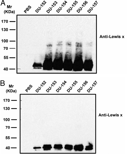
Fig. 5.
Immunoblot analysis of H. pylori-infected Capan 1 cells with mouse monoclonal anti-Lex antigen. Mock-transfected Capan 1 and Capan 1-FUCA2 K.D. cells were infected with different H. pylori strains (from DU-152 to DU-157) that were clinical isolates from 6 different patients with duodenal ulcer (DU). PBS represents a negative control in the absence of H. pylori. The coculture was maintained for 12 h at an MOI of approximately 400:1. In the parallel experiments, mock-transfected Capan 1 (A) and Capan 1-FUCA2 K.D. cells (B) were infected under the same conditions. The bacterial cells were collected and lysed for the Lex analysis. Lex-containing glycoproteins were found to greatly increase in the H. pylori cells co-cultured with mock-transfected Capan 1 cells (A), in contrast to those in the H. pylori cells co-cultured with Capan 1-FUCA2 K.D. cells (B).

