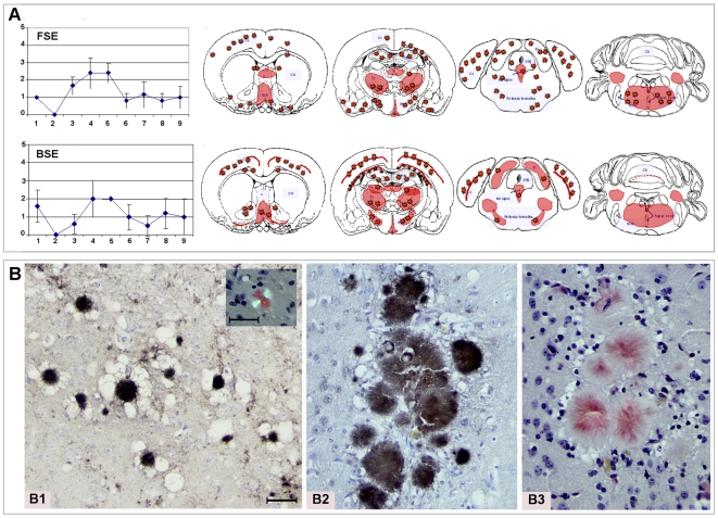
Figure 2. Similar FSE and BSE histopathological data in the TgOvPrP4 mice.
A. Similar FSE and BSE lesion profiles and PrPd mapping, observed in the brain of Tg(OvPrP4) mice (n = 5 to 6) infected with either cattle BSE or cheetah FSE at second passage. The nine gray matter sites of the lesion profiles were: 1. dorsal medulla nuclei, 2. cerebellar cortex, 3. superior colliculus, 4. hypothalamus, 5. central thalamus, 6. hippocampus, 7. lateral septal nuclei, 8. cerebral cortex at the level of thalamus, 9. cerebral cortex at the level of septal nuclei. The red color stands for the schematic representation of PrPd within the 4 brain levels analyzed. The red stars symbolize the florid plaque type of PrPd deposition. B.Typical florid plaques similarly detected in FSE and BSE transmission studies. Neuropathology of Tg(OvPrP4) mice inoculated with FSE agent from cheetah (case 1) (B1) or with BSE agent from cattle (B2 and B3) revealed the presence of typical florid plaques at first and second passage. Amyloid florid plaques sometimes formed clusters as in the cortex (B1 and B2: PrPd immunohistochemistry, B3 and insert: Red Congo staining). In these structures, the diameter of the florid plaques ranged from 30 to 100 µm.

