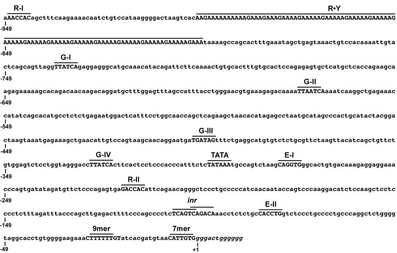
Figure 1.
Organization of the Dβ2 sequence analyzed for 5′ promoter activity. The positions of putative binding sites for Runx (R), GATA (G), and E proteins (E) are shown, as well as consensus TATA and initiator elements and polypurine·polypyrimidine DNA (R·Y). Dβ2 coding segments (italics) and 5′RSS sequences are also indicated. Numbering is relative to the first base of the Dβ2 coding sequence (+1).

