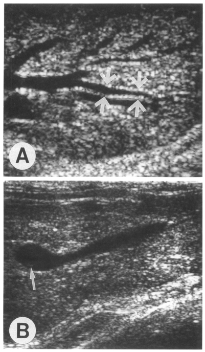
Fig. 1.
Sonograms of a rabbit (ID, 500-1) 9 weeks after infection with 500 metacercariae of Clonorchis sinensis. A. Transverse scan of the left hepatic lobe shows severe dilatation of the intrahepatic ducts (arrows). Note the hyperechoic bands along the duct wall, representing periductal fibrosis. B. Oblique scan of the gallbladder shows a few small echogenic foci (arrow) possibly indicating worms or desquamated materials.

