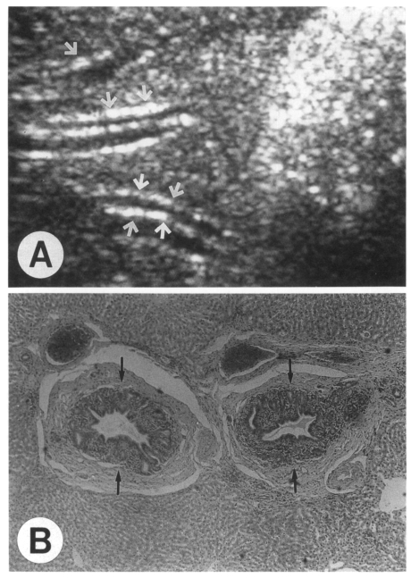
Fig. 2.
A rabbit (ID, 500-5) 18 months after treatment. A. Sonogram still shows moderate dilatation of the peripheral intrahepatic ducts with increased periductal echogenicity (arrows). B. Photomicrograph shows moderately persisted dilatation of the intrahepatic ducts and mucosal hyperplasia (arrows). Also note remaining periductal fibrosis which has been least resolved.

