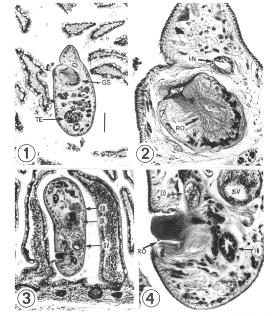
Figs. 1-4.
Figs. 1-2 Micrographs showing a longitudinally sectioned worm (worm A) in the small intestine of the patient. Fig. 1. A round genital sucker is located at the anterior 1/3 of the body. H & E stain. ×100. Fig. 2. Along the inner side of the genital sucker, there were 62 chitinous rodlets on the gonotyl arranged ovally. H & E stain. ×450.
Figs. 3-4. Micrographs showing a horizontally sectioned worm (worm C) in the small intestine of the patient. Fig. 3. The gonotyl (arrowhead) is protruded ventrally. The seminal receptacle and seminal vesicle are sectioned well. H & E stain. ×100. Fig. 4. A rodlet is seen on the gonotyl. H & E stain. ×450. ED; ejaculatory duct, GS; genital sucker, IN; intestine, RO; rodlet, SR; seminal receptacle, SV; seminal vesicle. TE; testis.

