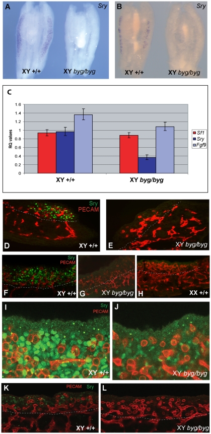
Figure 5. Loss of Sry transcript and protein expression in XY byg/byg gonads at 11.5 dpc on C57BL/6J.
(A) At the 17 ts stage, Sry transcript is detected throughout an XY control gonad (left) but is absent from the C57BL/6J XY byg/byg gonad (right). (B) By 19 ts, the signal is diminishing in the XY control gonad (left) and is still not detectable in the XY byg/byg gonad (right). (C) qRT-PCR analysis of Sry, Sf-1, and Fgf9 transcription in XY +/+ and byg/byg gonads at 11.5 dpc (17–18 ts). Error bars for the relative quantitation (RQ) values represent variation across four biological replicates for each genotype and three technical replicates for each sample. The 2.8-fold reduction in Sry levels in XY byg/byg gonads is significant (p = 0.02) based on a t-test calculated using average dCT values for the above genes and those of Hprt1. Differences in the levels of Sf-1 (p = 0.28) and Fgf9 (p = 0.23) were not significant, although there is a trend for reduced expression of Fgf9 in byg/byg. (D) Immunostaining of a longitudinal section of control XY gonad at 17 ts with anti-SRY (green) and anti-PECAM (red) antibodies reveals abundant expression of SRY in somatic cell nuclei of the gonad. Tissue beneath the dotted white line in this and subsequent images is mesonephric. (E) In contrast, very few SRY-positive cells are detectable in a longitudinal section from a XY byg/byg gonad at the same stage. (F) Confocal imaging of a control XY gonad after wholemount immunostaining with anti-SRY and anti-PECAM antibodies reveals large numbers of SRY-positive somatic cells, in contrast to XY byg/byg (G) and control XX (H) gonads. (I) High magnification confocal image of wild-type gonad at 11.5 dpc showing large numbers of SRY-positive somatic cells (green) and germ cells (red). (J) Confocal image of SRY expression in XY byg/byg gonad at 11.5 dpc generated using the same settings as in (I). Note the greatly reduced number of SRY-positive cells and the reduction in signal intensity in the mutant gonad. (K) A wild-type XY gonad at 11.0 dpc (13 ts) showing SRY-positive cells (green) amongst germ cells (red). (L) In contrast, no SRY-positive cells are detected at 11.0 dpc in an XY byg/byg gonad.

