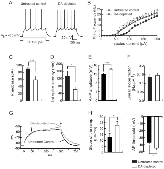
Figure 2. Dopamine depletion increases intrinsic excitability of medium spiny neurons.
(A) Representatives traces showing that evoked action potentials (120 pA current pulse) were increased in dopamine-depleted MSN. (B) Summary plot of discharge frequency as a function of injected current illustrating the effect of dopamine depletion on the firing frequency. Dopamine depletion significantly shifted the curve to the left. (C) Histogram of the mean rheobase values showing the decrease of the average minimal depolarizing current amplitude required to elicit spike discharge (rheobase) in dopamine-depleted MSN compared to untreated controls. (D) Summary histogram of the mean first spike latency at the 120 pA current pulse illustrating the decrease of the first spike latency induced by dopamine depletion. (E) Histogram illustrating the effect of dopamine depletion on AHP amplitude at the 120 pA current pulse. (F) Histogram values of mean linear slope of the current-frequency plot in both conditions. (G) Representatives traces from untreated control and dopamine-depleted conditions, show the slowly developing ramp potential preceding firing. The slope of the ramp membrane potential was estimated between 100 and 500 ms after the induction of the 500 ms-depolarising step (arrows). (H) The increase of the slope of the ramp in dopamine-depleted condition is illustrated in the summary histogram. (I) Histogram values of the action potential threshold in dopamine-depleted MSN compared to untreated controls. (Untreated control medium spiny neurons n = 8, dopamine (DA)-depleted medium spiny neurons n = 8; data represent mean ± SEM; *p<0.05, **p<0.01, ***p<0.001).

