
Fig. 2.
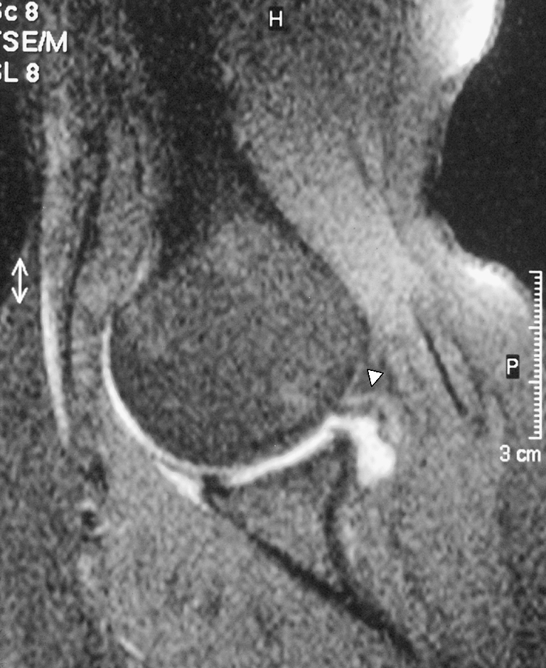
Arthroscopically proven partial-thickness supraspinatus tendon tear, type A. T1-weighted fat suppressed turbo spin echo (TSE) MR arthrography image in an oblique axial plane with the arm positioned in abduction and exorotation (ABER), obtained in a 40-year-old man, shows an example of a type A horizontal partial-thickness tear (arrow) without articular surface abnormality of the supraspinatus tendon