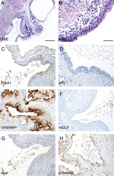
Fig. 2.
Tissue recombination experiment of Tgfbr2fspKO UGM with adult bladder epithelia. Bladder epithelia from wild-type C57Bl/6 mice were combined with UGM from Tgfbr2fspKO mouse embryo into collagen gel plug and grafted under the renal capsule of synergistic mice for 1 month. The histology of the resulting grafts was visualized by H&E staining at (A) 200× and (B) 400× magnification. Immunohistochemistry for the (C) endoderm marker FoxA1, (D) basal cell marker p63, (E) bladder urothelial marker uroplakin, (F) differentiated prostate epithelial markers mDLP secreted protein, and (G) PAP is shown in representative pictures as indicated. (H) Immunohistochemical staining for phosphorylated Smad2 indicated TGF-β signaling in the epithelial compartment, diminished in the stromal cells. The immunohistochemically stained sections were counterstained with hematoxylin (blue). Scale bar represents 200 μm (A) and 25 μm (B-H).

