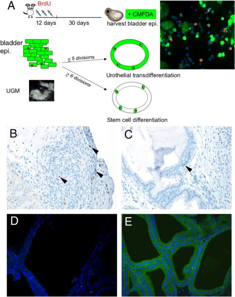
Fig. 6.
Tissue recombination of Tgfbr2floxE2/floxE2 UGM with labeled adult bladder epithelia. (A) The diagram illustrates the strategy for identifying BrdU label-retaining cells by the injection of BrdU in wild-type donor mice for 12 consecutive days, then harvesting the bladder 30 days later. Following the isolation of the bladder epithelia, the cells were further labeled with CMFDA. Dual labeling was confirmed by cytospin and fluorescence detection. The panel on the right illustrates urothelial cells immunostained for BrdU in red and green fluorescence of CMFDA. Two outcomes of the tissue recombinants with the labeled bladder epithelia could be observed: (1) If majority of the resulting prostatic epithelium in the grafts maintained detectable green CMFDA-labeled dye, it would suggest urothelial transdifferentiation as only a few cell divisions would be required (p5 cell divisions). (2) Alternatively, if only a few green cells were present maintaining BrdU (red), then it is likely that many more cell divisions (≥6) would be required, suggesting bladder stem cell induction as the primary means of prostate differentiation. Immunohistochemistry was used to detect BrdU in tissue recombinants after 1 month of grafting for (B) the control graft with bladder epithelial cells only (no mesenchyme) and (C) in the tissue recombinants with both bladder epithelia and UGM. Arrowheads indicate cells positive for BrdU staining. (D) Fluorescence microscopy of the UGM+urothelium tissue recombinant grafts showed little autofluorescence when the control epithelia were not labeled with CMFDA. (E) However, tissue recombinants with bladder epithelia pre-labeled with CMFDA showed nearly all epithelial cells were green 1 month after grafting. The tissue recombinants with UGM and bladder epithelial grafts in panels (C-E) had prostatic histology.

