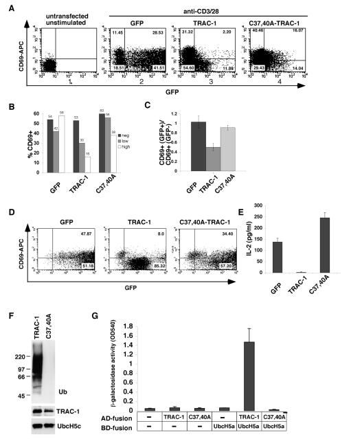
Figure 1. A TRAC-1/GFP fusion protein inhibits T cell activation.
A. Human primary T cells were nucleofected with peGFP-N3 (GFP, panel 2), or with peGFP-N3 containing TRAC-1 (panel 3) or C37,40A-TRAC-1 (panel 4) cDNA to express C-terminally tagged GFP fusion proteins. 16 h after transfection, cells were stimulated with plate-bound anti-CD3/28 for 18 h. Untransfected, unstimulated cells were included as controls (panel 1). Cells were stained with anti-CD69-APC, acquired by FACS and gated on live cells. The percentage of cells in each quadrant is indicated.
B. The percentage of CD69+ cells was calculated separately for GFP-negative (fluorescence intensity 0-10), GFP-low (fluorescence intensity 10-100) and GFP-high (fluorescence intensity >100) populations of the experiment in (A).
C. Six experiments were performed as in A. For each transfection, the percentage of CD69+ cells in the GFP+ population was divided by that in the GFP− population (see materials and methods). The averages of these ratios are represented for each of the transfected constructs.
D. Jurkat T cells were transfected with GFP, TRAC-1/GFP or C37,40A-TRAC-1/GFP expressing constructs. 24 h after transfection, GFP+ cells were sorted by MoFlo® and stimulated with plate-bound anti-CD3/CD28 for 48 h. FACS plots of cells stained with anti-CD69-APC are shown.
E. The culture supernatants of sorted Jurkat cells in (D) were analyzed for the presence of IL-2 by ELISA and represented in a graph (right). A representative of one out of two experiments is shown.
F. Bacterially expressed GST fusion proteins of TRAC-1 and C37,40A-TRAC-1 were captured on glutathione-sepharose beads and incubated for 90 min at 30°C with purified E1, His-tagged E2 UbcH5c, ubiquitin and ATP. After the reaction, beads were analyzed by Western blotting with antibodies against ubiquitin to detect ubiquitin ligase activity (top). The reaction mixture was blotted with anti-GST (middle) and anti-His (bottom) to detect the presence of TRAC-1 proteins and UbcH5c, respectively.
G. Yeast was transformed with GAL4 binding domain (BD) and activation domain (AD) containing vectors, either without insert (-) or with TRAC-1 (wildtype or the C37,40A mutant) or UbcH5a as inserts, as indicated. An interaction between proteins results in β-galactosidase activity and was measured with CPRG as substrate.

