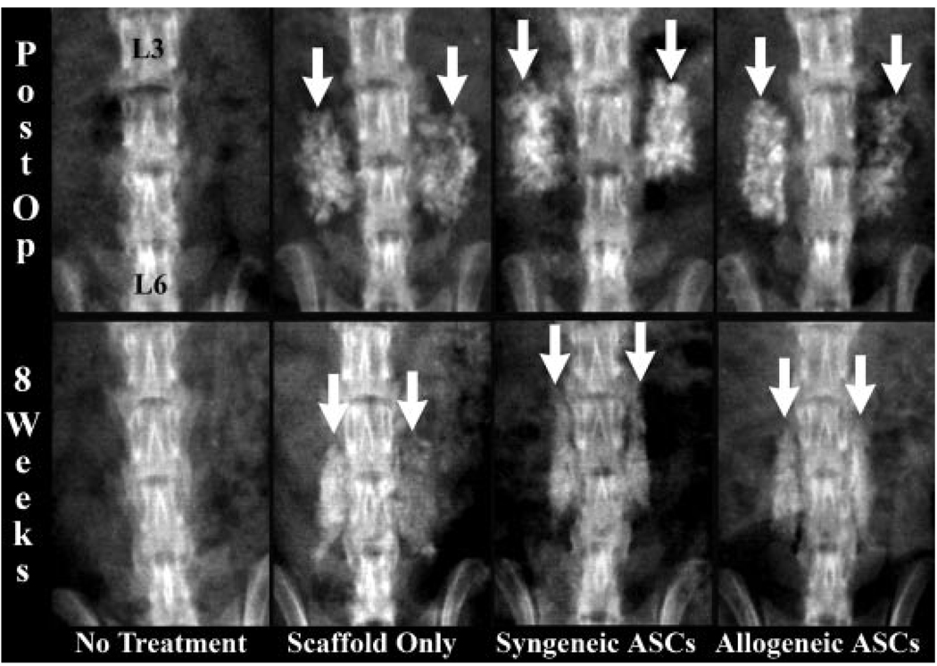
Figure 2.
Radiographs of the lumbar vertebra performed immediately (postop) and 8 weeks after implantation (8 weeks) of representative animals in each treatment cohort. The lumbar vertebra labeled in the first image are congruent with all images shown. The radioopaque scaffold is evident in the postop radiographs from scaffold treatment cohorts (white arrows). Callus is smaller, more organized, and less active in the ASC treatment cohorts 8 weeks after implantation (white arrows).

