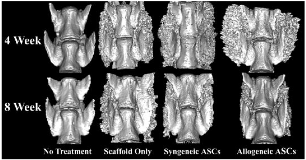
Figure 3.
Representative 3D reconstructions from µ-CT slices of the L4 (top) and L5 (bottom) vertebral bodies viewed from the anterior surface of the four treatment cohorts at each time point. Callus was more highly organized in the spines with ASC loaded scaffolds versus those with scaffold alone 8 weeks after implantation, consistent with a higher degree of remodeling.

